Imaging
The Imaging Unit (IU) of the NKUA Core Facility has been established at the premises of the School of Sciences, NKUA. The preparatory phase of different samples viewing has started in Autumn 2024 and it is expected that the IU will be in full operation capacity by early summer 2025. We aim to provide state-of-the-art high-resolution light and electron microscope imaging, serving the needs of all NKUA schools and beyond. Currently, our team consists of three members, all with high experience and expertise in Imaging techniques, including Electron Microscopy (sample preparation and viewing). At this phase of its operation the unit is divided at two sub-units:
- Transmission Electron Microscope unit (TEM)
- Scanning Electron Microscope unit (SEM)
Staff
Head of Unit

Professor Ioannis Trougakos
Imaging Unit
Co-Head of Unit

Professor Nikolaos Kavantzas
Imaging unit
Principal Investigator

Dr. Georgios Baltatzis
Imaging Unit
TRANSMISSION ELECTRON MICROSCOPE (TEM)
SERVICES
Transmission Electron Microscopy (TEM) is an essential tool for the detailed examination of thin sections at high magnification. It allows for ultra-high-resolution imaging at magnifications up to 600,000X, providing crucial insights into cellular and subcellular structures but also in other preparations (if compatible). Routine TEM observation ensures accurate evaluation of tissues/cells fine structure by detecting ultrastructural changes that are not visible under light microscopy.
TECHNOLOGY AND EQUIPMENT
Hitachi Transmission Electron Microscope (TEM) ΗΤ7800
- Transmission Electron Microscopy (TEM) is an essential tool for the detailed examination of thin sections at high magnification. It allows for ultra-high-resolution imaging at magnifications up to 600,000X, providing crucial insights into cellular and subcellular structures but also in other preparations (if compatible). Routine TEM observation ensures accurate evaluation of tissues/cells fine structure by detecting ultrastructural changes that are not visible under light microscopy.
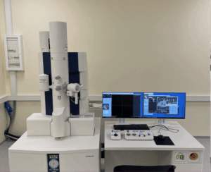
Hitachi Transmission Electron Microscope (TEM) ΗΤ7800, capable of performing sample tomography and 3D reconstruction.
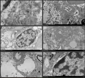
Routine TEM analysis for renal biopsies(ultra-high-resolution imaging for medical diagnostics)
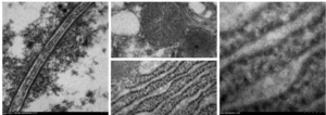
TEM high magnification
]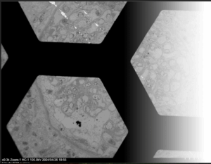
Sample panoramic images
Scanning Electron Microscope (SEM)
SERVICES
Scanning Electron Microscopy (SEM) is a powerful technique for examining the surface morphology of various specimens, including fresh organic material or biological samples (e.g., Drosophila). SEM provides high-resolution, three-dimensional imaging that reveals intricate surface structures from any type of inorganic samples( g., rocks, minerals, etc.) at magnifications ranging from low to ultra-high resolution.
TECHNOLOGY AND EQUIPMENT
Hitachi Scanning Electron Microscope SU3800
- Scanning Electron Microscopy (SEM) is a powerful technique for examining the surface morphology of various specimens, including fresh organic material or biological samples (e.g., Drosophila). SEM provides high-resolution, three-dimensional imaging that reveals intricate surface structures from any type of inorganic samples( g., rocks, minerals, etc.) at magnifications ranging from low to ultra-high resolution.
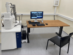
Hitachi Scanning Electron Microscope (SEM) SU3800
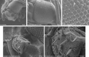
Fresh organic material – Drosophila samples (SEM images)
The IU is also equipped with the following supportive instruments from Leica:
- UC7 Ultramicrotome
- UC7 Cryo-Ultramicrotome, with an integrated liquid nitrogen cooling system (FC7), for cryo-electron microscopy samples
- EM TRIM 2 Pyramid Trimmer
- EM KMR3 Glass Knife Maker for preparing thin sections of samples
- EM AC20 Electron Microscopy Sample Staining Device
- EM TP Tissue Processor for sample preparation in Electron Microscopy
- EM FSP Freeze Substitution System
- EM CPD300 Critical Point Dryer for drying samples for scanning electron microscopy
- EM ACE200 Sputter Coater and Carbon Coater for preparing samples for scanning electron microscopy

Book our Equipment
We look forward to supporting your research with the best tools and expertise available. Explore our resources and unlock new possibilities at NKUA Core Facilities today!
You can use your NKUA account to book our equipment quickly and easily by clicking the button below.
If you are an external user, please use the appropriate booking form.
Availability
External Users
Book our Equipment
We look forward to supporting your research with the best tools and expertise available. Explore our resources and unlock new possibilities at NKUA Core Facilities today!
You can use your NKUA account to book our equipment quickly and easily by clicking the button below.
If you are an external user, please use the appropriate booking form.